 Ultrasound is a specialized diagnostic imaging procedure, where sound waves are used to visualize the internal structures of the body. It is painless, does not involve radiation, and can usually be done without any anaesthesia or sedation.
Although ultrasound may be used on many areas of the body, the most common sites for ultrasound in veterinary medicine include the abdomen and the heart. Abdominal ultrasound can give us a detailed look at the liver, pancreas, adrenal glands, stomach, intestines, lymph nodes, spleen, and urinary bladder. If necessary, biopsies can often be obtained with a needle guided by ultrasound, avoiding the need for more invasive surgery.
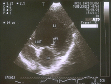
Echocardiography, or cardiac ultrasound, can give us a detailed view of the heart as it is beating. We can see and measure the sizes of the different chambers in the heart, assess the heart valves, and determine how smoothly and quickly blood is flowing through the heart. Echocardiography is an extremely useful and necessary tool when diagnosing the cause of a heart murmur, and determining the cause and extent of heart disease.
The value and accuracy of an ultrasound is completely determined by the ability and experience of the person performing the ultrasound. At Yorkville Animal Hospital, we can arrange for ultrasounds to be performed by a board certified internal medicine specialist right here in our hospital. By using the services of a specialist that does this procedure all the time, we can ensure that our clients are receiving the very best care possible for their pet.